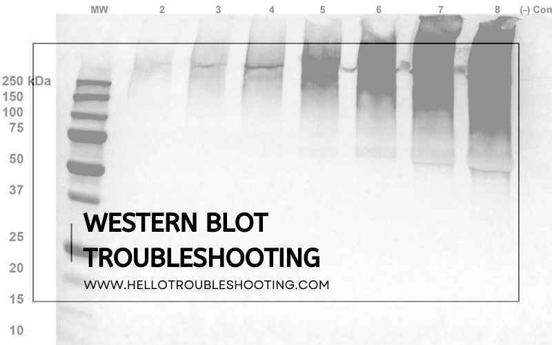
Ensure antibodies are fresh and properly stored. Verify protein transfer efficiency and correct blocking conditions for optimal results.
Western blot troubleshooting can be challenging but is crucial for accurate results. Common issues include weak signals, high background, and non-specific bands. Addressing these problems requires a systematic approach. Fresh antibodies and proper storage maintain their activity. Verifying protein transfer efficiency ensures proteins move correctly from gel to membrane.
Correct blocking conditions reduce non-specific binding, enhancing signal clarity. Regularly checking reagents and equipment prevents many common issues. Proper technique and attention to detail can significantly improve western blot outcomes. Understanding these key factors helps researchers obtain reliable and reproducible data. This guide offers essential tips for effective troubleshooting, ensuring successful western blot experiments.

Introduction To Western Blotting
Western blotting is a powerful technique in molecular biology. It helps detect specific proteins in a sample. Scientists use it to study protein expression and modifications.
Purpose And Applications
Western blotting serves many purposes in research. It helps confirm protein identity and quantity. This technique is vital in diagnosing diseases. Researchers use it to study gene expression patterns. It plays a crucial role in validating other experiments.
Basic Principles
The process begins with sample preparation. Proteins are extracted from cells or tissues. These proteins are then separated using gel electrophoresis. A polyacrylamide gel helps in this separation.
Next, the proteins are transferred to a membrane. This membrane is usually made of nitrocellulose or PVDF. After transfer, the membrane is blocked to prevent non-specific binding.
Antibodies are then used to detect the target protein. The primary antibody binds to the protein of interest. A secondary antibody, linked to an enzyme or dye, binds to the primary antibody. This allows visualization of the protein on the membrane.

Credit: m.youtube.com
Common Issues In Western Blotting
Western blotting is a powerful method for detecting proteins. Sometimes, things can go wrong. Understanding common issues can save time and effort.
Poor Signal
Poor signal in Western blotting can be frustrating. There are several causes:
- Insufficient protein loading: Ensure enough protein is loaded.
- Antibody concentration: Use optimized antibody concentrations.
- Transfer issues: Verify efficient protein transfer to the membrane.
Check the following table for quick fixes:
| Issue | Possible Fix |
|---|---|
| Low protein amount | Load more protein |
| Weak antibody binding | Increase antibody concentration |
| Poor transfer | Optimize transfer conditions |
High Background
High background can obscure results. It often results from:
- Non-specific binding: Block the membrane properly.
- Excess antibody: Reduce antibody concentrations.
- Inadequate washing: Increase washing steps.
Consider these steps to reduce high background:
- Use blocking agents like BSA or milk.
- Optimize primary and secondary antibody dilutions.
- Extend washing times with TBST.
Sample Preparation Tips
Effective sample preparation is key for successful Western Blot results. This section covers essential tips to ensure high-quality samples. Follow these guidelines to improve your Western Blot outcomes.
Lysate Quality
Quality lysates are critical for reliable Western Blot results. Here are some tips:
- Cell Lysis: Use appropriate lysis buffer for your samples.
- Avoid Degradation: Add protease and phosphatase inhibitors.
- Keep Cold: Perform lysis on ice to prevent protein degradation.
Protein Quantification
Accurate protein quantification ensures consistent loading. Follow these steps:
- Use a Reliable Assay: Choose a BCA or Bradford assay.
- Standard Curve: Always run a standard curve with your samples.
- Replicates: Perform quantification in triplicates for accuracy.
| Step | Description |
|---|---|
| 1. Cell Lysis | Use appropriate lysis buffer and keep samples cold. |
| 2. Inhibitors | Add protease and phosphatase inhibitors. |
| 3. Quantification | Use a reliable assay and perform in triplicates. |
These tips can greatly enhance your Western Blot results. Ensure high lysate quality and accurate protein quantification for better outcomes.
Gel Electrophoresis Optimization
Optimizing gel electrophoresis is crucial for successful Western Blot results. This step ensures clear and distinct protein bands. Read on to master gel selection and running conditions.
Choosing The Right Gel
Choosing the correct gel type is essential. It depends on the protein size.
- Polyacrylamide Gel: Suitable for proteins between 5-200 kDa.
- Agarose Gel: Ideal for larger proteins or nucleic acids.
Also, pay attention to the gel percentage. It affects the resolution of protein bands.
| Gel Percentage | Protein Size Range (kDa) |
|---|---|
| 6% | 50-200 |
| 10% | 20-100 |
| 15% | 10-50 |
Selecting the correct gel ensures better separation and clearer bands.
Running Conditions
Running conditions impact the quality of your results. Proper voltage and time are key.
- Voltage: Use 100V for stacking gel and 150V for resolving gel.
- Time: Run for 60-90 minutes, depending on gel type and protein size.
Also, the buffer system matters. Commonly used buffers include Tris-Glycine and Bis-Tris.
Always monitor the progress. Adjust conditions if bands appear smeared or distorted.
Following these tips will help you achieve sharp, well-resolved bands.
Transfer Efficiency Improvement
Achieving optimal transfer efficiency is crucial for Western blot success. This process ensures proteins move from gel to membrane effectively. Enhanced transfer efficiency leads to clearer, more accurate results. Below, we explore methods to improve this crucial step.
Membrane Selection
Choosing the right membrane is essential. There are two main types: nitrocellulose and PVDF. Both have unique qualities that affect transfer efficiency.
| Membrane Type | Advantages | Disadvantages |
|---|---|---|
| Nitrocellulose |
|
|
| PVDF |
|
|
Transfer Methods
Different transfer methods can impact efficiency. Two common methods are wet transfer and semi-dry transfer.
- Wet Transfer: This method uses a tank filled with transfer buffer. It provides high transfer efficiency, especially for larger proteins. A downside is the longer transfer time.
- Semi-Dry Transfer: This method is faster and uses less buffer. It’s ideal for smaller proteins but may have lower efficiency for larger proteins.
To improve efficiency, ensure proper setup of the transfer apparatus. Use fresh buffers and avoid air bubbles in the setup.
Blocking And Antibody Incubation
Western blotting is a powerful technique for detecting specific proteins. However, troubleshooting is often necessary. One critical stage is Blocking and Antibody Incubation. Proper blocking and antibody incubation ensure clear, specific bands and minimize background noise.
Blocking Buffers
Blocking buffers prevent non-specific binding of antibodies. This reduces background noise in your blot.
Common blocking buffers include:
- Bovine Serum Albumin (BSA): Effective for many antibodies. Use at 1-5% concentration.
- Non-fat Dry Milk: Cost-effective and works for most antibodies. Use at 3-5% concentration.
- Casein: Preferred for phospho-specific antibodies. Use at 0.5-1% concentration.
Choose a buffer that suits your specific antibodies and proteins. Also, consider the compatibility with your detection system.
Primary And Secondary Antibodies
Primary antibodies bind to your target protein. Secondary antibodies bind to the primary, amplifying the signal.
Tips for using antibodies:
- Primary Antibody: Optimize concentration through titration. Too much or too little affects results.
- Secondary Antibody: Use at recommended dilution. Ensure it matches the host species of the primary antibody.
- Incubation Times: Longer incubation increases sensitivity. Shorter times reduce background.
Proper antibody handling is crucial. Store antibodies at recommended temperatures and avoid repeated freeze-thaw cycles.
Here’s a quick reference table for antibody incubation:
| Antibody | Concentration | Incubation Time |
|---|---|---|
| Primary | 0.5-2 µg/mL | 1-2 hours at room temp or overnight at 4°C |
| Secondary | 1:1000 – 1:5000 | 1 hour at room temp |
By paying close attention to blocking and antibody incubation, you can achieve clear, specific results in your Western blot experiments.
Detection Techniques
Western Blotting is a crucial technique in molecular biology. Detection techniques are key to identifying specific proteins. The two main methods are Chemiluminescence and Fluorescence. Each has unique advantages and limitations.
Chemiluminescence
Chemiluminescence is a popular detection method for Western Blot. This technique uses a substrate that emits light when it reacts with an enzyme. The light signal is then captured on film or a digital camera.
- Advantages:
- High sensitivity
- Easy to use
- Can detect low-abundance proteins
- Limitations:
- Short signal duration
- Requires special equipment
- Quantification can be tricky
To improve results, optimize the enzyme and substrate concentrations. Also, use high-quality antibodies for better signal clarity.
Fluorescence
Fluorescence detection is another effective method. This technique uses fluorescently-labeled antibodies. The signal is detected using a fluorescence scanner or imager.
| Advantages | Limitations |
|---|---|
|
|
Ensure proper handling of fluorescent antibodies to maintain signal strength. Minimize exposure to light to reduce photobleaching.
Data Analysis And Interpretation
Data analysis and interpretation are critical in Western Blot experiments. Properly analyzing your data ensures accurate and reliable results. You must understand how to quantify bands and apply normalization techniques.
Quantifying Bands
Quantifying bands involves measuring the intensity of protein bands on your blot. You can use software tools designed for this purpose. These tools help you obtain precise data from your experiment.
Here’s a simple way to quantify bands:
- Scan your blot using a high-resolution scanner.
- Upload the image to your analysis software.
- Select each band and measure its intensity.
Ensure your bands are well-separated to avoid overlap. Overlapping bands can lead to inaccurate measurements. Use a molecular weight marker to identify the bands precisely.
Normalization Techniques
Normalization techniques ensure your data is accurate and comparable. They help correct for loading and transfer variations. The most common method is using housekeeping proteins.
Follow these steps for normalization:
- Select a stable housekeeping protein. Examples include GAPDH or β-actin.
- Measure the intensity of the housekeeping protein band.
- Divide the intensity of your target protein by the housekeeping protein intensity.
This ratio gives you the normalized value. Always verify that your housekeeping protein remains constant across samples. This ensures your normalization is valid.
Another method is using total protein normalization. This technique involves staining the entire membrane and measuring the total protein load.
Here’s a comparison table for the two methods:
| Method | Advantages | Disadvantages |
|---|---|---|
| Housekeeping Protein | Simple, widely used | Housekeeping protein must be stable |
| Total Protein | More accurate | Requires additional staining |
Choose the method that best suits your experiment’s needs. Consistent normalization ensures your data is reliable and valid.
Troubleshooting Guide
Welcome to our Western Blot Troubleshooting Guide. This section helps you solve common problems with Western Blotting. Follow these tips to get the best results in your experiments.
Faint Bands
Faint bands can be frustrating. Here are some steps to fix this issue:
- Check Protein Load: Ensure you have loaded enough protein. Use a protein ladder to compare.
- Antibody Concentration: Use the correct concentration of primary and secondary antibodies. Too little can lead to faint bands.
- Incubation Time: Increase the incubation time with your primary antibody.
- Detection Reagents: Ensure your detection reagents are fresh and properly mixed.
Non-specific Bands
Non-specific bands can make your results unclear. Consider these tips:
- Blocking Solution: Use a proper blocking solution to reduce background noise. Common solutions include BSA or non-fat milk.
- Antibody Specificity: Ensure your primary antibody is specific to your target protein.
- Washing Steps: Increase the number and duration of washing steps to remove non-specific binding.
- Secondary Antibody: Use a secondary antibody that is highly specific and validated.
| Problem | Possible Cause | Solution |
|---|---|---|
| Faint Bands | Low protein load | Increase protein concentration |
| Non-Specific Bands | Inadequate blocking | Use proper blocking solution |
Follow these tips to troubleshoot your Western Blot issues. With the right approach, you can achieve clear and reliable results.
Expert Tips And Best Practices
Western Blotting is a crucial technique in molecular biology. To achieve reliable results, one must follow expert tips and best practices. Below, we share key insights to enhance your Western Blot troubleshooting skills.
Documentation
Proper documentation is essential in Western Blot troubleshooting. Keep detailed notes of every step. This includes:
- Sample preparation: Record the source and concentration of samples.
- Electrophoresis conditions: Note the voltage and duration.
- Transfer conditions: Document the type of membrane and transfer buffer used.
- Antibody details: List the dilution and incubation times.
Use a table for quick reference:
| Step | Details |
|---|---|
| Sample Preparation | Source, concentration |
| Electrophoresis | Voltage, duration |
| Transfer | Membrane type, buffer |
| Antibody | Dilution, incubation |
Reproducibility
Reproducibility ensures that your results are reliable. Follow these steps:
- Standardize protocols: Use the same reagents and conditions.
- Control samples: Include positive and negative controls in every run.
- Replicate experiments: Perform each experiment at least three times.
- Consistent environment: Maintain consistent temperature and humidity.
These practices help in minimizing variations. They also enhance the credibility of your results.
Frequently Asked Questions
Why Is My Western Blot Showing No Bands?
Your primary antibody might be too dilute. Check concentration and incubation times to ensure optimal binding.
How Can I Reduce Background Noise?
Use a blocking buffer with the right concentration. Also, ensure thorough washing steps to remove excess antibodies.
Why Are My Bands Smeared?
Protein degradation or overloading the gel can cause smearing. Ensure fresh samples and appropriate loading amounts.
What Causes Uneven Band Intensity?
Uneven gel loading or transfer can lead to this issue. Use consistent loading techniques and ensure even transfer.
How Do I Prevent Non-specific Binding?
Use highly specific primary and secondary antibodies. Implement stringent washing steps to remove non-specific proteins.
Conclusion
Effective Western blot troubleshooting ensures accurate and reliable results. By following these tips, you can overcome common challenges. Regularly check your reagents, equipment, and protocols. Stay patient and methodical in your approach. Consistency is key to mastering Western blot techniques.
Happy experimenting and may your results always be clear and precise.
